An in silico testbed for fast and accurate MR labeling of orthopedic implants
- Electrical & Computer Eng. Dept, Worcester Polytechnic Institute, United States
- Ansys, United States
- Dassault Systèmes Deutschland GmbH, Germany
- Neva Electromagnetics, LLC, United States
- GE HealthCare, United States
- Micro Systems Enigineering, Inc, an affiliate of Biotronik, United States
- Musculoskeletal Translational Innovation Initiative, Department of Orthopedic Surgery, Beth Israel Deaconess Medical Center and Harvard Medical School, United States
- Harvard Medical School, United States
- Athinoula A Martinos Center for Biomed. Imaging, Massachusetts General Hospital, United States
Figures
Figure 1
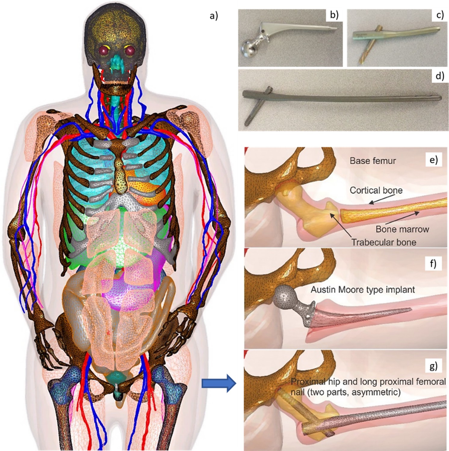
Visible Human Project (VHP)-Female Model with Embedded Passive Implants.
Left (a) – surface CAD meshes for the Visible Human Project (VHP)-Female model (with some muscles removed for purposes of visualization); and right – examples of passive femoral implants embedded into the model. (b–d) At top right – physical femoral implants; (e–g) at center and bottom – anatomically justified CAD realization within the virtual human VHP-Female. An Austin Moore implant is shown in b; a short proximal femoral nail with the proximal hip (a large femoral neck screw) is given in c; a long proximal femoral nail with the proximal hip is presented in d.
Figure 2
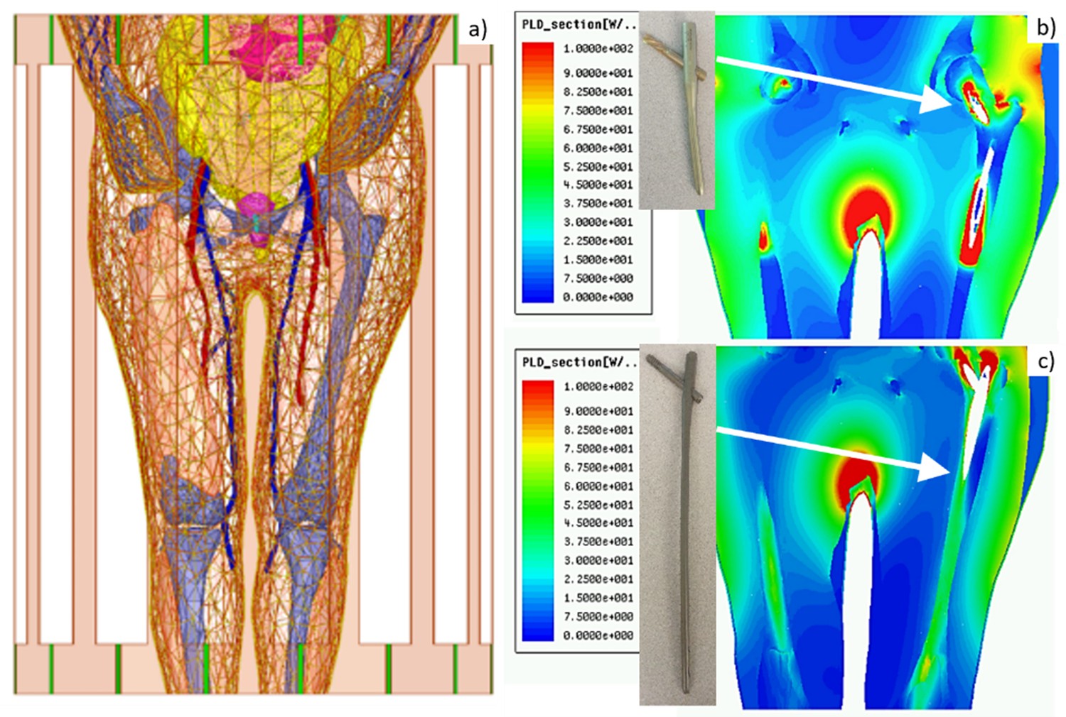
Left – Visible Human Project (VHP)-Female computational phantom positioned within a 1.5 T MRI birdcage coil at the abdominal landmark.
Right – power loss density in W/m3 in the coil for the (b) Austin Moore and (c) femoral nail implants.
Figure 3
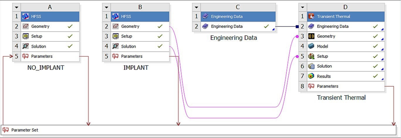
Ansys Workbench modeling workflow consisting of the HFSS (electromagnetic) module labeled as A and B, and the transient thermal module labeled D.
Thermal material properties are contained in module C.
Figure 4
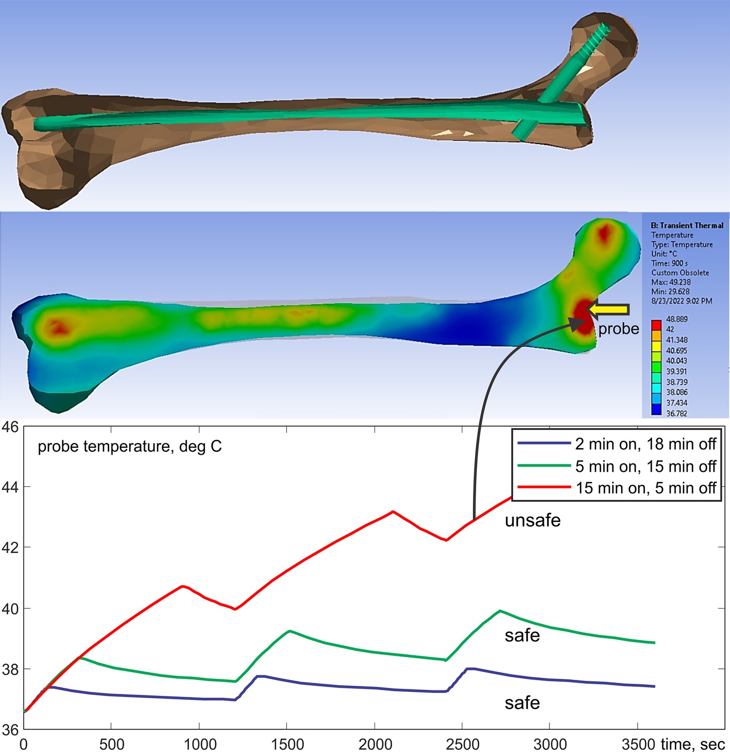
Top – long femoral nail subject to three repetitions of 15 min exposure followed by 5 min of rest for 1 hr in total.
Center – temperature contour plot in a cut plane roughly bisecting the embedded femoral implant at the end of the last heating cycle. Bottom – temperature rise profile at the temperature probe. Only the bone is shown but the computations are performed for the entire model.
Figure 5
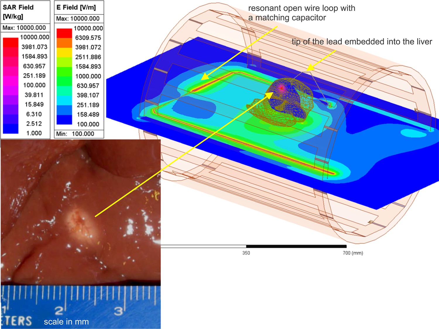
Liver experiment setup: E-field distribution in the plane of the resonant loop and specific absorption rate (SAR) distribution within liver in a plane passing through the tip of the lead.
Bottom left – lesion in a section of ex vivo bovine liver created with heating at the tip of a 16 gauge (1.3 mm) bare copper wire needle in about 1.5 min (Hue et al., 2018).
Additional files
Download links
A two-part list of links to download the article, or parts of the article, in various formats.
Downloads (link to download the article as PDF)
Open citations (links to open the citations from this article in various online reference manager services)
Cite this article (links to download the citations from this article in formats compatible with various reference manager tools)
An in silico testbed for fast and accurate MR labeling of orthopedic implants
eLife 12:RP90440.
https://doi.org/10.7554/eLife.90440.3


